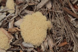
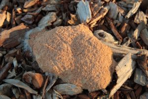
Ceratiomyxa fruticulosa – P Vallier
What are slime moulds?
Slime moulds are classified as Protista (or Protocista). They are neither plants, animals nor fungi. Slime moulds are peculiar protists that normally take the form of amoeba but also develop fruit bodies that release spores, and are superficially similar to the sporangia of fungi.
One of the earliest references to slime moulds is in The Garden of Earthly Delights by Hieronymus Bosch (c. 1504), in which 22 slime moulds are depicted. There are almost 1000 species worldwide, and they occupy nearly every habitat on earth (primarily moist terrestrial). They eat bacteria, fungi and decaying organic matter. Amazingly, they seem to have an ability to communicate and organize themselves, and can solve maze puzzles.
There are two types of Slime Moulds:
- Cellular Slime Moulds are rarely observed in their vegetative state because of their small size. They spend most of their life as single-celled amoeboid protists, but upon the release of a chemical signal, the individual cells aggregate into a great swarm (known as a pseudoplasmodium) and eventually into multicellular slugs.
- Plasmodial Slime Moulds are easier to observe in nature.
Life Cycle of Plasmodial Slime Moulds
The plasmodium is a single cell bound only by a thin, elastic membrane. It has multiple nuclei and is often pigmented. At this stage the slime mould feeds on decaying organic matter, bacteria, protozoa and other minute organisms by actively engulfing the small particles and by absorbing soluble food directly into the mass. The stage is capable of locomotion, and a rapid flow of cytoplasm can readily be seen in the vein-like regions of the plasmodium.
In unfavourable conditions the plasmodium may revert to a dormant hardened mass. When the weather gets too cold or dry, the plasmodium hardens into a sclerotium. The sclerotium consists of irregular hardened masses of large cysts (macrocysts), each containing a number of nuclei. In this dormant form, the slime mould is viable for a period of years and often survives winter in this state.
When conditions are right and/or the food supply is exhausted, the mature plasmodium begins spore formation (sporulation). The plasmodium moves to an exposed, drier portion of the substrate, and begins the formation of fruit bodies (eg. Sporangia). The fruit bodies produce spores that are subsequently dispersed in the environment.
The spores of slime moulds are nearly always round. They range from smooth, finely warty, echinate or spiny, roughened to coarsely reticulated. The spores are typically fairly large (9-15um, but some are as small as 5um).
Spores germinate to form either amoeboid cells (myxamoebae) or flagellated swarm cells. These can revert to a dormant micvrocyst when conditions are unfavourable. (In favourable conditions, fusion of cells occurs to produce a single cell zygote, which feeds and grows to form the plasmodium.)
There are four types of fruit bodies:
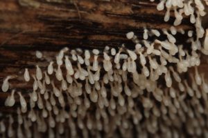
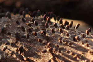
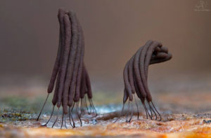
- The Sporangium is the most common type. It is a small spore container, sessile or stalked, with wide variations in colour and shape. Typical components of a sporangium, useful for identification, are a peridium (outer wall), spores, a capillitium (delicate network of hair-like elements), a columnella or pseudocolumella, a stalk and a hypothallus (thin membranous structure at the base). Sporangia usually occur in groups, since they form from separate portions of the same plasmodium.
Examples are Hemitrichia calyculata, which looks a little like a coffee-coloured ice-cream cone. Trichia favoginea, appearing somewhat similar to honeycomb, Arcyria denudata, resembling pink fairy floss. Stemonitis splendens, and Ceratiomyxa fruticulosa.
- The Aethalium is cushion-shaped, sessile and relatively large.
Examples include Lycogala epidendrum, which grows on wood and is commonly called ‘Wolf’s milk’, and Fuligo septica, often seen on wood chip mulch and sometimes called ‘Dog’s Vomit’.
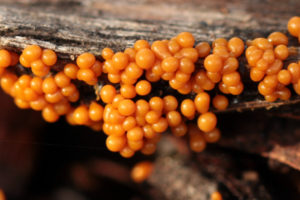
- The Pseudoaethalium is composed of sporangia closely crowded together, and is usually sessile, although a few may be stalked.
An example is Tubifera ferruginosa, which takes the form of attractive bright coral-pink ‘cakes’.
- The Plasmodiocarp is usually sessile. Plasmodiocarps take the form of the plasmodial veins from which they were derived.
An example is Hemitrica serpula, which usually a yellow mustard colour. The Plasmodium coalesces to form a network of swollen veins.
References:
Lloyd, Sarah, Where the slime mould creeps, Edition. 2 Tympanocryptis Press, Birralee Tasmania. See here for a review by Paul George and here to purchase this book from Fungimap Shop
Stephenson, S.L. & Stempen, H (1994). Myxomycetes: a handbook of slime molds. Timber Press, Portland, Oregon
Baumann, K., Like Nothing On Earth, the Incredible Life Of Slime Moulds, (video)
This is a summary of an illustrated talk given by Paul George to the Fungi Group of the Field Naturalists Club of Victoria 7 November 2005. The report is written by Virgil Hubregtse
By Sarah Lloyd for her hands-on workshop in Tasmania on slime moulds for National Science Week Aug 2018
‘Slime Moulds – Nature’s miniature jewels’.
Acellular or plasmodial slime moulds – the myxomycetes – have baffled scientists and naturalists for centuries. They have been placed in the kingdoms plant, fungi, animal and protista, based on their very different life stages comprising single-celled amebae, moving feeding plasmodia and spore-bearing ‘fruits’. They are now considered to be amoebozoans.
Myxamoebae – the first feeding stage of a myxomycete – can comprise up to 50% of soil microbes. They feed on bacteria and other single-celled organisms and are important in recycling nutrients and enriching soils. Their second feeding plasmodial stage is occasionally visible on logs, stumps and soil. During favourable conditions plasmodia start to produce fruiting bodies.
Because slime moulds are ephemeral and extremely small, they are easily overlooked. They are among the least studied of all the microorganisms and Australia is believed to be the least studied country in the world. Temperate forests are known to be the richest sites for slime moulds so living in the middle of a Tasmanian forest is an ideal place to study them.
In 2010 Sarah Lloyd started to search regularly for slime moulds in the forest surrounding her home. Fruiting bodies or plasmodia often emerge overnight or in the morning and many are brightly coloured and highly visible when they first appear. Their location can be marked and their progress monitored as they mature and become less conspicuous. Fruiting bodies are collected in good condition (i.e. before they are spoiled by rain, invertebrates or fungi) and lodged at the National Herbarium of Victoria where they are available for study.
Tasmania’s wintry weather is perfect for slime mould activity. As the weather changes from cold and frosty to wild, windy and wet, plasmodia creep about among leaf litter and over logs and stumps, eventually to form fruiting bodies on leaves, twigs and wood.


